

Digital radiology may represent the greatest technological advancement in medical imaging in the last decade. The use of radiographic films in X ray imaging might become obsolete in a few years. An appropriate analogy that is easy to understand is the replacement of typical film cameras with digital cameras. Images can be immediately acquired, deleted, modified, and subsequently sent to a network of computers.
In 1995, the world celebrated the 100th anniversary of the discovery of x-rays by W.C. Röntgen. His discovery was the first to enable the display of human internal anatomical structures and it revolutionized the field of medicine. Since then, the use of x-rays has contributed to the diagnosis and treatment of many diseases thereby helping to improve the health of people all over the world. Medical imaging systems have developed from simple units used to image specific anatomical sites to systems that can visualize the whole body, obtain information concerning functional aspects of specific organs and even yield information about the chemistry taking place in organs and tissues. 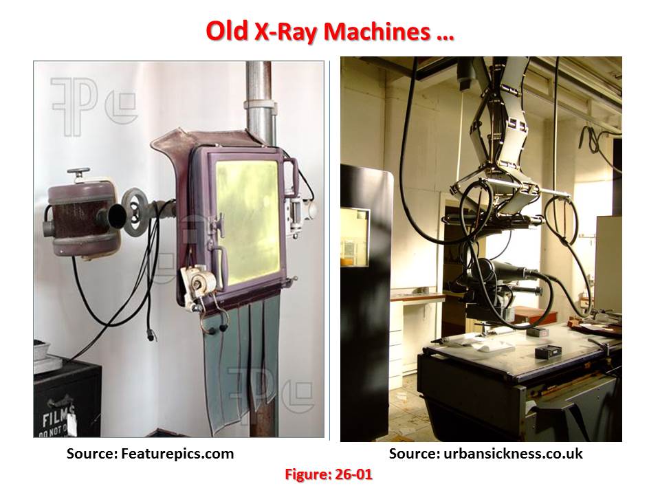 Imaging with x- rays utilizes a range of techniques. Projection radiography using screen–film technology is the most common imaging modality used in radiology. The x-rays are attenuated in the body depending on the type and the thickness of the tissues through which they have to pass. The transmitted x-rays are detected by special screens emitting light after interaction with x-rays. The emitted light is further detected by the film. Dual screen, dual emulsion film systems are commonly used in radiographic procedures such as chest, abdominal and skull radiography. When high resolution is needed, as in mammography, single screen and single emulsion film systems are used. During the radiography examination, the x-ray attenuation information about the human anatomy is projected into the two dimensions of the radiograph. Once the exposed film is chemically processed to create a visible image, it can be displayed on a light box, transported wherever it is needed and kept as an archival record. X-ray film systems enable the radiologist to acquire, display, communicate and store image data in a simple way. This technique remains one of the most widely used medical diagnostic tools.
Fluoroscopy is an imaging procedure that allows real time viewing of anatomical structures. As in projection radiography, this is a two dimensional imaging technique but it uses image intensifiers and displays images on a TV monitor. Fluoroscopic systems have undergone significant technological advances in recent years. For example, they are available with image intensifiers allowing the viewing of the entire abdomen. Depending on the application, these systems are available in a wide variety of modifications (Gastrointestinal, Angiographic, etc.). Hospitals usually have suites dedicated to specialized applications. The long fluoroscopy times, especially during interventional procedures, may result in local patient doses that cause deterministic effects of radiation in patients. It is important to optimize such investigations to reduce unnecessary exposure of patients and medical staff.
A basic problem in imaging with x-rays (or other penetrating radiation) is that a two-dimensional image is obtained of a three-dimensional object. This means that structures can overlap in the final image, even though they are completely separate in the object. This is particularly troublesome in medical diagnosis simply because there are many anatomic structures that can interfere with what the physician is trying to see. During the 1930’s, this problem was attacked by moving the x-ray source and detector in a coordinated motion during image formation. From the geometry of this motion, a single plane within the patient remains in focus, while structures outside this plane become blurred. This is analogous to a camera being focused on an object at 5 feet, while objects at a distance of 1 and 50 feet are blurry. These related techniques based on motion blurring are now collectively called classical tomography. The word tomography means “a picture of a plane.”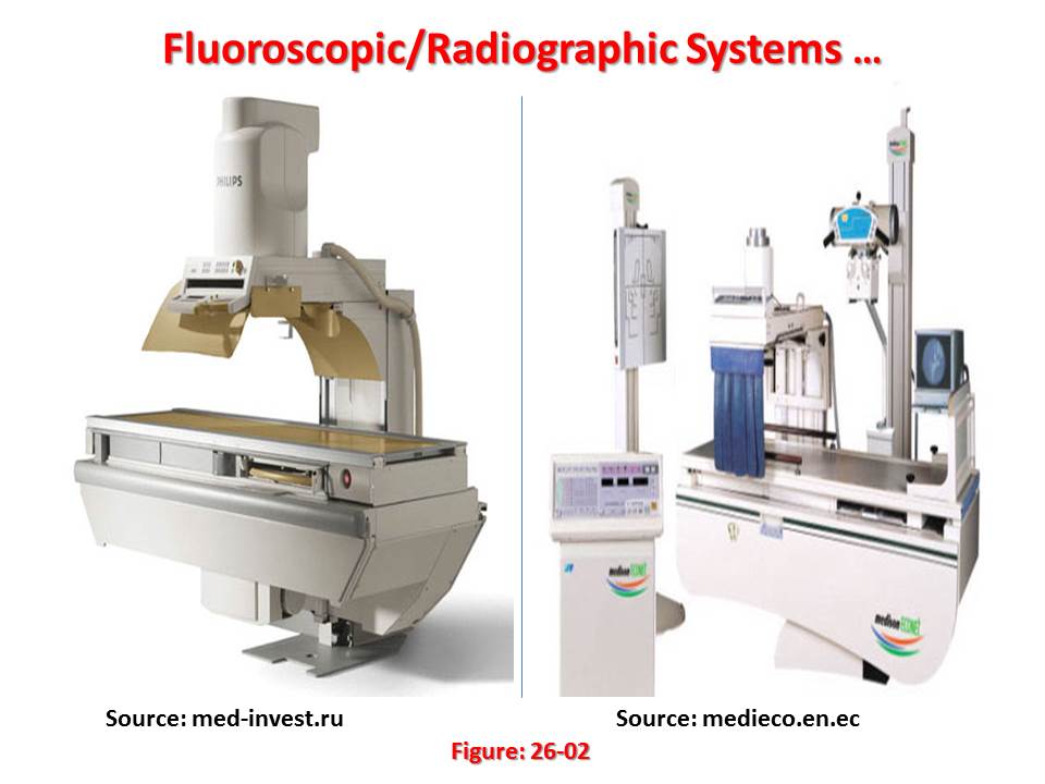 In spite of being well developed for more than 50 years, classical tomography is rarely used. This is because it has a significant limitation: The interfering objects are not removed from the image, only blurred. The resulting image quality is usually too poor to be of practical use. The long sought solution was a system that could create an image representing a 2D slice through a 3D object with no interference from other structures in the 3D object.
This problem was solved in the early 1970s with the introduction of a technique called Computed Tomography (CT). CT revolutionized the medical x-ray field with its unprecedented ability to visualize the anatomic structure of the body. CT was originally introduced to the marketplace under the names Computed Axial Tomography and CAT scanner. Continuing technological developments, such as spiral (or helical) scanning and multislice scanners, have improved both the speed and quality with which images are obtained. Fast scan times and rapid image reconstruction have allowed the development of real time CT scanning. Development of CT was only made possible by a massive development in computer technology. CT started the era of computerized diagnostic methods that are gradually replacing traditional film based diagnostic techniques.
CT also represents one of the earliest forms of digital x-ray imaging, in which images are captured and stored in a digital format. More recent developments in digital radiology include digital fluorography employing an image intensifier and computed radiography utilizing a special storage phosphor plate which retains the latent image. Unlike computed radiography, direct digital radiography uses an active matrix detector, typically of amorphous selenium or a phosphor coupled to silicon to convert X ray energy to a digital signal virtually instantaneously with no intermediate operational steps. Digital imaging provides key advantages in the manipulation, storage and transmission of images.
Advances in imaging have facilitated the development of interventional radiological procedures, in which imaging is used to help guide therapeutic procedures. For example, angioplasty is done fluoroscopically and involves placing and expanding a balloon catheter inside a blood vessel so as to dilate the vessel and improve blood flow. Interventional radiology continues to evolve and other commonly used therapeutic techniques make use of imaging technologies. In this Code of Practice, separate descriptions of methodology are given for the areas of general radiography, fluoroscopy, mammography, CT and dental radiography. In this context, general radiography implies all radiography apart from the latter four modalities.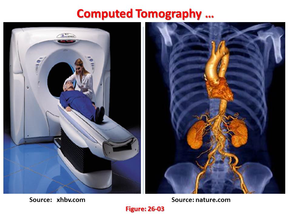 Nowadays, medical imaging equipment is taking advantage of modern digital technology and has become a symbol of ‘high technology’.
Digital radiological techniques offer the potential for improved image quality and, given the higher sensitivity of its image receptors compared with film, also offers the potential for dose reduction. In practice, however, since image receptors also have a broader dynamic range than film, higher doses are correspondingly possible.
American engineers began developing digital technology in the mid-twentieth century. Their techniques were based on mathematical concepts suggested by the seventeenth-century German mathematician, Gottfried Wilhelm Leibniz, who proposed a binary computing system. His innovation inspired such numerical codes as American Standard Code for Information Interchange (ASCII) that described objects with digits.
Digital technology is a base two process. Digitized information is recorded in binary code of combinations of the digits 0 and 1, also called bits, which represent words and images. Digital technology enables immense amounts of information to be compressed on small storage devices that can be easily preserved and transported. Digitization also quickens data transmission speeds. Digital technology has transformed how people communicate, learn, and work.
Telecommunications has relied on digital methods to transmit messages. In the early 1980s, enhanced fiber optics enabled the development of digital communication networks. Digital technology replaced analog signals for many telecommunication forms, particularly cellular telephone and cable systems. Analog-to-digital converters utilized pulse code modulation (PCM) to change analog data into digital signals. Compared to analog transmissions, digitized signals were less distorted and could easily be duplicated.
In the early 2000s, digital computers ranging from laptops to Internet networks came in many sizes and performed various tasks. Supercomputers performed complex mathematical calculations analyzing vast amounts of data. The Digital Data Broadcast System (DDBS) guided air-traffic control. Digital radiography converted analog signals of x-rays to create digital images. Digital information was stored on plastic disks with pitted patterns of 1s and 0s that lasers translated. By the early 2000s, digital cameras had transformed photography by recording color and light intensities with pixels. In addition, Joint Photographic Experts Group (JPEG) and the Moving Picture Experts Group (MPEG) codes achieved digital compression of images and video. Animation had often been digitized with some films and cartoons being created entirely with computers.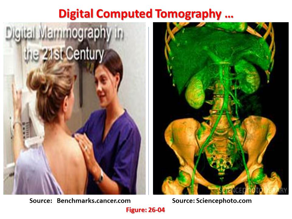 Digital imaging techniques came to x-ray in the 1980s when analog to digital (A/D) converters and computers were also adapted to conventional fluoroscopic image intensifier/TV systems. Digital imaging has led to a similar improvement and renaissance in x-ray just as it has in home and professional audio. Just as many recordings and music albums now sound sharper and better than ever on a digital compact disk (CD) player than on and older (analog) record player, digital x-rays often look sharper and cleaner than the analog version. Many of the fluoroscopic (“fluoro” for short) x-ray procedures described have benefited greatly from the addition of digital technology. Further, angiographic procedures for looking at the blood vessels in the brain, kidneys, arms and legs, and the blood vessels of the heart all have benefited tremendously from the adaptation of digital technology.
It is envisaged that over the next ten to fifteen years a large majority of conventional (basic) x-ray systems will also be upgraded to all digital technology. In addition to digital fluoroscopic systems, eventually, digital x-ray detectors will replace the majority of film cassette/film screen systems. This technology is currently very new and is now available at a handful of sites worldwide. An intermediate step called phosphor plate technology is currently available at hundreds of sites around the world. These plates trap the x-ray energy and require an intermediate processing step to release the stored information so it can be converted into a digital picture.
Benefits of digital technology to all x-ray systems include:
- Lower dosage x-rays can often be used to achieve the same high quality picture as with film;
- Digital x-ray images can be enhanced and manipulated with computers and sent via a network to other workstations and computer monitors so that many people can share the information and assist in the diagnosis;
- Digital images can be archived onto compact optical disks or digital tape drives saving tremendously on storage space and manpower needed for a traditional x-ray film library
- Digital images may be retrieved from an electronic archive for future reference.
Some modalities like mammography require extremely high-resolution film to show the small breast cancers and calcifications. Digital detectors capable of a similarly high resolution are under developments that expect to be available in the future. However, digital imaging is already being used in parallel to high-resolution film in breast imaging and breast biopsy systems.
The electro-medical equipment market is divided into two digital imaging market segments: The diagnostic market and the therapeutic market.
The diagnostic imaging equipment market represents the portion of medical equipment that enables diagnosis by producing images of a chosen area or organ of the body. The market comprises five main sectors:
- X-ray Imaging;
- Ultrasound;
- Magnetic Resonance Imaging (MRI);
- Computed Tomography (CT); and
- Nuclear Medicine.
Over recent decades, alternative medical imaging methods have been developed. These methods use x-rays in a different way, for instance CT, use other sources of radiation (Nuclear Medicine), or do not rely on ionizing radiation at all (Ultrasound and MRI). Nevertheless, x-ray imaging continues to be used as the first imaging modality by medical imaging professionals, and it holds more than 50 percent of the diagnosis market. Projection x-ray imaging is divided into the following sub segments:
- General Radiography;
- Radiography & fluoroscopy (R&F);
- Angiography;
- Cardiovascular Mammography; and
- Dental (Intra-oral and Panoramic).
The reality is that according to an International Atomic Energy Agency’s report, digital technology advances have facilitated an increase in the application of CT. The use of rapid wide-area multiple-slice CT, for example, has expanded the use of CT to a large range of applications from cardiology to paediatric investigations. Such new technology brings with it increasing radiation doses and challenges the established practices of dose determination. Radiological diagnosis is an area of medicine that is vital to effective health care. On average, every second person in the world has a radiological examination each year.
This report also shows that the number of x-ray radiological examinations has more than doubled in the last 20 years (Data based on the United Nations Scientific Committee on the Effects of Atomic Radiation). There is a marked imbalance in the geographical distribution of services — in effect, less than 2 percent of the total examinations conducted worldwide are in low income countries. Another distinctive current feature of radiology is the rate of technological change, characterized by a dramatic move from analogue images, such as film, to digital imaging techniques.
The conclusion of the report was that for low income countries, digital technology brings with it unexpected opportunities, as well as challenges. Unfortunately, many developing countries still rely almost completely on manual development of film to produce images for diagnosis. This methodology is technically challenging, often resulting in poor quality images. It is also environmentally unfriendly. Especially critical, however, is the way in which such processing can limit effective service delivery, where radiological equipment and skilled manpower are scarce. Digitized medical images can be sent electronically to distant places, allowing remote or resource limited locations to access centres of excellence for expert diagnosis and to assist in professional training. With the maturing of this technology and further cost reductions, digital imaging appears increasingly financially viable in developing countries. The continuation of improvements in digital technology promises a future alternative to the manual processing of film images, bringing with it the hope of a more efficient and widespread usage of radiological services.
Resources:
- Dosimetry in Diagnostic Radiology – An International Code of Practice;
- The Scientist and Engineer’s Guide to Digital Signal Processing;
- Encyclopedia;
- Answers;
- Answers – Quicken;
- Answers – Distorted;
- Imaging;
- IDS Healthcare; and
- IAEA – Nuclear Technology Review 2010.
- This chapter was published on “Inuitech – Intuitech Technologies for Sustainability” on May 26, 2012; and
- This chapter was updated on 26 June 2020.